The Gram stain, the most widely used staining procedure in bacteriology, is a complex and differential staining
Is a Gram stain a direct stain?
The Gram stain is a direct method, since the cells themselves retain dye. In indirect, or negative, staining, smears are produced by mixing material with India ink or acidic dyes such as nigrosine.
Is Gram stain a special stain?
A Gram stain of a skin lesion is a laboratory test that uses special stains to detect and identify bacteria in a sample from the skin. The Gram stain method is one of the most commonly used techniques to quickly diagnose bacterial infections.
Is Gram stain a simple stain?
The Gram stain is a differential stain, as opposed to the simple stain which uses 1 dye. As a result of the use of 2 dyes, making this procedure a differential stain, bacteria will either become purple/blue or pink during the procedure.Why is Gram staining called a differential staining?
It is called a differential stain since it differentiates between Gram-positive and Gram-negative bacteria. Bacteria that stain purple with the Gram staining procedure are termed Gram-positive; those that stain pink are said to be Gram-negative. … Chemically, 60 to 90% of the Gram-positive cell wall is peptidoglycan.
What are the types of staining in microbiology?
A variety of staining techniques can be used with light microscopy, including Gram staining, acid-fast staining, capsule staining, endospore staining, and flagella staining.
What is Gram stain reaction?
A Gram stain is a test that checks for bacteria at the site of a suspected infection such as the throat, lungs, genitals, or in skin wounds. … When the stain combines with bacteria in a sample, the bacteria will either stay purple or turn pink or red. If the bacteria stays purple, they are Gram-positive.
What is the secondary stain in Gram staining?
Gram stain permits the separation of all bacteria into two large groups, those which retain the primary dye (gram-positive) and those that take the color of the counterstain (gram-negative). The primary dye is crystal violet and the secondary dye is safranin O.Is Endospore stain a differential stain?
The endospore stain is a differential stain used to visualize bacterial endospores. Endospores are formed by a few genera of bacteria, such as Bacillus . By forming spores, bacteria can survive in hostile conditions. Spores are resistant to heat, dessication, chemicals, and radiation.
What type of stain is the Gram stain and what does it rely on for meaningful results?Some labels will NOT be used. What type of stain is the Gram stain, and what does it rely on for meaningful results? A. It is a simple stain that relies on chemical differences in the plasma membrane to yield meaningful results.
Article first time published onWhat is Gram stain morphology?
The Gram stain test has three components – colony morphology, Gram reaction (purple and red) and microscopic morphology. Colony morphology is a description of how the microorganism looks when it grows on an agar plate. It includes color, shape and other characteristics of the colony.
What is gram staining an example of?
Gram stain or Gram staining, also called Gram’s method, is a method of staining used to classify bacterial species into two large groups: Gram-positive bacteria and Gram-negative bacteria. The name comes from the Danish bacteriologist Hans Christian Gram, who developed the technique in 1884.
What is gram staining quizlet?
Gram stain technique. A staining procedure used to identify bacterial cells as gram-positive or gram-negative. developed by christian gram in the 1800s. -Cells are stained with crystal violet and Gram iodine solution and washed with a decolorizer. -Safranin is applied as a counterstain.
What are the types of staining?
- Oil Stain. Oil stains are the most widely available and the type of stain most people think of when they think of stain. …
- Varnish Stain. Varnish stains resemble oil stains in every way but one. …
- Gel Stain. …
- Lacquer Stain. …
- Water-Soluble Dye Stain. …
- Metal-Complex (Metalized) Dye Stain.
What is staining and its types?
The types are: 1. Simple Staining 2. … Gram Staining 4. Acid Fast Staining 5. Endospore Staining.
Which stain is a differential stain?
One commonly recognizable use of differential staining is the Gram stain. Gram staining uses two dyes: Crystal violet and Fuchsin or Safranin (the counterstain) to differentiate between Gram-positive bacteria (large Peptidoglycan layer on outer surface of cell) and Gram-negative bacteria.
What color will a Gram-positive cell stain?
Gram-positive cells have a thick peptidoglycan layer and stain blue to purple.
What Gram stain is Streptococcus?
Streptococci are Gram-positive, nonmotile, nonsporeforming, catalase-negative cocci that occur in pairs or chains.
What is Endospore staining in microbiology?
Endospore staining is a technique used in bacteriology to identify the presence of endospores in a bacterial sample. Within bacteria, endospores are protective structures used to survive extreme conditions, including high temperatures making them highly resistant to chemicals.
What is a stain in microbiology?
staining. [stān´ing] artificial coloration of a substance to facilitate examination of tissues, microorganisms, or other cells under the microscope.
How are staining techniques classified?
How are stains classified? Stains are classified based on the pH of their chromophore (color bearing ion) into acidic, basic and neutral. Acidic dyes have anionic chromophore eg., sodium+ eosinate-. Basic dyes have cationic chromophore eg., methylene blue+ chloride-.
What is stain chemical?
Rather than dying the fibers (as with aniline dyes) or putting fine pigment particles on the surface (as with conventional “stains”) a chemical stain reacts with the natural tannin in the wood to produce a brown to reddish brown. The depth of the color that can be achieved is stunning.
What color are endospores after a Gram stain?
After gram staining, the endospore is colorless. After the endospore stain, it is green.
What color is a negative endospore stain?
positive/negative: endospores stain green and may be seen inside cells or outside cells. endospore negative cells stain red with no evidence of green staining.
What color do endospores and bacteria stain in an endospore stain?
Whereas the counterstain (safranin) is pink/reddish in color, the primary stain (malachite green) is green in color. Therefore, endospores will appear green in color while the vegetative cells will pink/reddish in color under the microscope.
What is a secondary stain?
First cells are stained with crystal violet, followed by the addition of a setting agent for the stain (iodine). Then alcohol is applied, which selectively removes the stain from only the Gram negative cells. Finally, a secondary stain, safranin, is added, which counterstains the decolorized cells pink.
What is the primary and secondary stains for the Endospore stain?
It is the most widely used technique for endospore staining. … The method utilizes malachite green as the primary stain and safranin as counterstain.
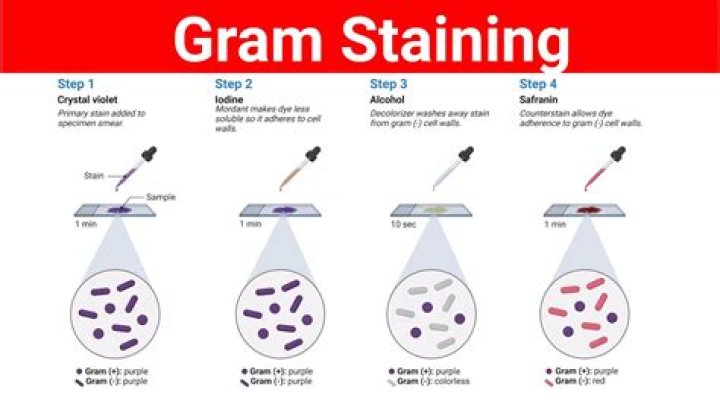
What is the order of Gram staining?
The stains are applied to a smear of bacteria on a microscope slide in the following order: crystal violet, Gram’s iodine, decolorizing agent, and safranin.
What do the Gram stain the acid fast stain and the Endospore stain have in common?
-Heat fix slide. What do the Gram stain, acid-fast stain, and endospore stain have in common? … They use heat to force the dye into cell structures. They are simple stains.
What is the role of iodine in Gram staining?
Gram’s iodine is used in Gram staining procedure to differentiate gram positive and gram negative organisms. Gram’s iodine acts as a mordant that causes the crystal violet to penetrate and adhere to the gram –positive organisms.
How do you do a Gram stain?
- Apply a smear of bacteria on to a slide. …
- Add about 5 drops of Hucker’s Crystal Violet to the culture. …
- Add about 5 drops of iodine solution to the culture. …
- Tilt slide and decolorize with solvent (acetone-alcohol solution) until purple color stops running. …
- Add about 5 drops of Safranine O.