Each lung is enclosed within a cavity that is surrounded by the pleura. The pleura (plural = pleurae
What are the lungs separated by?
The lungs are separated by the mediastinum. This area contains the heart, trachea, esophagus, and many lymph nodes. The lungs are covered by a protective membrane known as the pleura and are separated from the abdominal cavity by the muscular diaphragm.
Where are the lungs positioned?
The lungs are located on either side of the breastbone in the chest cavity and are divided into five main sections (lobes).
What is the area between the two lungs called?
The mediastinum is the partition between the lungs and includes the mediastinal pleura. It is commonly applied to the internal between the two pleural sacs, the sternum and the thoracic vertebral column extending to the diaphragm.What separates the lungs from the thoracic cavity quizlet?
A dome-shaped sheet of muscle attached to the thoracic wall that separates the lungs and thoracic cavity from the abdominal cavity. As the chest cavity enlarges, the diaphragm moves downward and flattens to create a vacuum that allows air to flow into the lungs.
Why lungs are divided into lobes?
The fissures are formed in early prenatal development by invaginations of the visceral pleura that divide the lobar bronchi, and section the lungs into lobes that helps in their expansion. The right lung is divided into three lobes by a horizontal fissure, and an oblique fissure.
What is the name of the membrane surrounding the lungs?
The pleura includes two thin layers of tissue that protect and cushion the lungs. The inner layer (visceral pleura) wraps around the lungs and is stuck so tightly to the lungs that it cannot be peeled off. The outer layer (parietal pleura) lines the inside of the chest wall.
What is the root of the lung?
Hila, or lung roots, are relatively complicated structures that consist mainly of the major bronchi and the pulmonary arteries and veins. The hilum of the lung is found on the medial aspect of each lung, and it is the only site of entrance or exit of structures associated with the lungs.What tubes bifurcate from the windpipe?
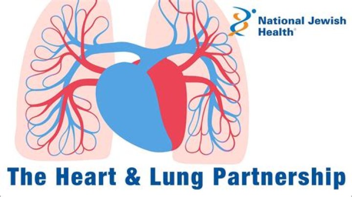
The trachea begins just under the larynx (voice box) and runs down behind the breastbone (sternum). The trachea then divides into two smaller tubes called bronchi: one bronchus for each lung.
What are the parts of lungs?Lungs. Bronchial tubes/bronchi. Bronchioles. Air sacs (alveoli)
Article first time published onHow are the two lungs different than one another?
You have two lungs, but they aren’t the same size the way your eyes or nostrils are. Instead, the lung on the left side of your body is a bit smaller than the lung on the right. This extra space on the left leaves room for your heart. Your lungs are protected by your rib cage, which is made up of 12 sets of ribs.
What separates the two lungs quizlet?
What separates the lungs? The lungs are separated by the mediastinum that contains the trachea, the heart and the great vessels.
What separates the two lungs how many lobes does each lung have?
Each lung is separated into lobes branching off the main bronchus; the right lung has three lobes, while the left has only two lobes. As the bronchi branch out, the total area of the two new branches is larger than its parent bronchus, making it extremely easy for the air to rush into the lungs.
What encloses the lungs and attaches them to the inside of the thoracic cavity?
The diaphragm is the flat, dome-shaped muscle located at the base of the lungs and thoracic cavity. The lungs are enclosed by the pleurae, which are attached to the mediastinum.
What is pleural separation?
The pleural cavity is the potential space between the pleurae of the pleural sac that surrounds each lung. … The serous membrane that covers the surface of the lung is the visceral pleura and is separated from the outer membrane the parietal pleura by just the film of pleural fluid in the pleural cavity.
What type of membrane surrounds each of the lungs quizlet?
The pleural membranes completely surround each lung.
What layer of a membrane is closest to the organ?
Serous membranes have two layers. The parietal layers of the membranes line the walls of the body cavity (pariet- refers to a cavity wall). The visceral layer of the membrane covers the organs (the viscera). Between the parietal and visceral layers is a very thin, fluid-filled serous space, or cavity.
What is the pleura?
(PLOOR-uh) A thin layer of tissue that covers the lungs and lines the interior wall of the chest cavity. It protects and cushions the lungs. This tissue secretes a small amount of fluid that acts as a lubricant, allowing the lungs to move smoothly in the chest cavity while breathing.
Where does the trachea split?
When the trachea reaches the lungs, it splits into two tubes: the right bronchus and the left bronchus. Each of these enters a lung. The trachea and bronchi share many structural components, including smooth muscle, cartilage, and cilia.
Which are two airways that split from the trachea and enter the lungs?
The trachea branches into two smaller airways: the left and right bronchi, which lead to the two lungs.
Where the trachea forks into each lung?
At chest-level, the trachea forks into the bronchi. The bronchi in turn spread out into narrower channels called the bronchial tubes. The bronchi carry oxygen to two large organs called the lungs, which are located inside the chest. Each lung is made up of roughly 300 million smaller sacks called pulmonary alveoli.
What structures enter and exit the hilum of the lung?
Anatomy of the Hilum The major bronchi, pulmonary arteries, pulmonary veins, and nerves are the structures which enter and exit the lungs in this region.
What does the hilum of the lung contain?
The hilum contains mostly bronchi and pulmonary vasculature, along with the phrenic nerve, lymphatics, nodes, and bronchial vessels. Both left and right hilum contain a pulmonary artery, pulmonary veins (superior and inferior), and bronchial arteries.
What is hilum in chest?
The hilum is what connects your lungs to their supporting structures and where pulmonary vessels enter and exit your lungs. … Each hilum is in a flat area at the center of each lung, toward your spine or the back of your lungs (medial surface).
Which of the following comprises the lungs?
QuestionWhich of the following comprises the lungs? a. Tracheae, b. 1∘bronchi, c. 2∘ bronchi, d. 3∘ bronchi, e. initial bronchioles, f. terminal bronchioles, g. duct of alveoli, h. alveoliChapter NameBreathing And Exchange Of GasesSubjectBiology (more Questions)Class11thType of AnswerVideo, Text & Image
What is the muscular sheet below the lungs called?
The diaphragm, located below the lungs, is the major muscle of respiration. It is a large, dome-shaped muscle that contracts rhythmically and continually, and most of the time, involuntarily. Upon inhalation, the diaphragm contracts and flattens and the chest cavity enlarges.
What is the significance of having only two lobes in the left lung whereas in the right lung It has three lobes?
The left lung has only two formal lobes because of the space taken up in the left side of the chest cavity by the heart, though it does have the lingula, which is similar to a lobe.
What is the function of spirometer?
Spirometer. A spirometer is a diagnostic device that measures the amount of air you’re able to breathe in and out and the time it takes you to exhale completely after you take a deep breath. A spirometry test requires you to breathe into a tube attached to a machine called a spirometer.
What is the name of the muscle that separates the lung cavity from the abdomen and helps regulate breathing quizlet?
The diaphragm is a flat, dome-shaped muscle below the lungs. It separates the thoracic cavity from the abdominal cavity.
How many fissures are in the right lung?
The right lung has two fissures, oblique fissure and horizontal fissure, which separate the lung into three lobes – upper, middle, and lower.
Where is the oblique fissure located?
The oblique fissure, which extends from the costal to the mediastinal surface of the lung both above and below the hilum. It divides the left lung in an upper and a lower lobe and in the right lung, separates the inferior from the middle and superior lobes, and is closely aligned with the fissure in the left lung.