The posterior cranial fossa is part of the cranial cavity
What cranial nerves are in the posterior fossa?
Some authors describe innervation of the posterior fossa dura by the facial and glossopharyngeal nerves,36 as well as from the hypoglossal and vagus nerves. The branch from the vagus starts from the superior ganglion, follows the posterior meningeal artery, and supplies the posterior fossa dura.
What drains from posterior cranial fossa?
Each petrosal vein drains not only the brainstem and the anterior aspect of the cerebellum, but also the neighboring cranial nerves. Venous injection of the posterior fossa shows, at least, one drainage vein for each one of cranial nerves V, VI, VII, and VIII. – Anterior cerebellar veins, superior and inferior.
What structures are supported by the posterior cranial fossa?
The posterior cranial fossa houses the brainstem and cerebellum. The brainstem is comprised of the medulla oblogata, pons and midbrain and continues down through the foramen magnum to become the spinal cord.What passes through cranial fossa?
- Summary.
- Olfactory Nerve (CN I)
- Optic Nerve (CN II)
- Oculomotor Nerve (CN III)
- Trochlear Nerve (CN IV)
- Trigeminal Nerve (CN V)
- Abducens Nerve (CN VI)
- Facial Nerve (CN VII)
What is the biggest foramen in the posterior cranial fossa and give the structure passes through it?
Foramen magnum: You can’t miss this one! The name in Latin means “great hole” and it has to be relatively large to allow the lower end of the brainstem and the upper end of the spinal cord to pass through. Importantly, the foramen magnum also allows the vertebral arteries to enter the skull.
Which parts of the brain are found in the anterior middle and posterior cranial fossa?
(b) The complex floor of the cranial cavity is formed by the frontal, ethmoid, sphenoid, temporal, and occipital bones. The lesser wing of the sphenoid bone separates the anterior and middle cranial fossae. The petrous ridge (petrous portion of temporal bone) separates the middle and posterior cranial fossae.
What are the 3 cranial fossa?
A cranial fossa is formed by the floor of the cranial cavity. There are three distinct cranial fossae: Anterior cranial fossa (fossa cranii anterior), housing the projecting frontal lobes of the brain. Middle cranial fossa (fossa cranii media), separated from the posterior fossa by the clivus and the petrous crest.What are the components of the brainstem select all that apply?
The three components of the brainstem are the medulla oblongata, midbrain, and pons. Brainstem Anatomy: Structures of the brainstem are depicted on these diagrams, including the midbrain, pons, medulla, basilar artery, and vertebral arteries.
What is in the anterior cranial fossa?The anterior cranial fossa is an important anatomical landmark in clinical orthodontics consisting of the frontal, ethmoid, and sphenoid bones. The relationships between these bones remain poorly understood.
Article first time published onWhat is fossa in anatomy?
Fossa – A shallow depression in the bone surface. Here it may receive another articulating bone or act to support brain structures. Examples include trochlear fossa, posterior, middle, and anterior cranial fossa.
What forms a roof over the posterior cranial fossa?
The tentorium, which is attached along the petrous ridges, roofs the posterior fossa.
What are the cranial cavity contents?
The cranial cavity houses the Brain, Meninges, and the Cerebrospinal Fluid.
Which opening is present in the middle cranial fossa?
The middle part of the fossa presents, in front, the chiasmatic groove and tuberculum sellae; the chiasmatic groove ends on either side at the optic foramen, which transmits the optic nerve and ophthalmic artery to the orbital cavity.
What passes through the foramen cecum?
The foramen cecum varies in size in different subjects, and is frequently impervious; when open, it transmits the emissary vein from the nose to the superior sagittal sinus.
Is the occipital lobe in the posterior fossa?
Posterior cranial fossa – This fossa is relatively deep and contains the cerebellum. Projecting posterior to the foramen magnum is the internal occipital protuberance. The internal acoustic meatus is located on the petrous portion of the temporal bone. The jugular foramen is anterolateral to the hypoglossal canal.
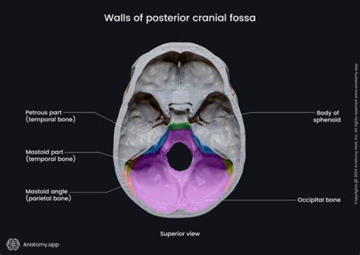
What bones contribute to the posterior cranial fossa and how is this fossa demarcated from the middle cranial fossa and the foramen magnum?
The boundaries of the posterior cranial fossa are formed anteriorly by the dorsum sellae, posterior aspects of the body of the sphenoid and the basilar part of occipital bone; posteriorly by the squamous part of the occipital bone; laterally by the petrous and mastoid parts of the temporal bone and by the lateral parts …
What are the 3 parts of the brainstem?
The brainstem is divided into three sections in humans: the midbrain (mesencephalon), the pons (metencephalon), and the medulla oblongata (myelencephalon).
What are the 3 parts of the brainstem and their functions?
The brainstem (brain stem) is the distal part of the brain that is made up of the midbrain, pons, and medulla oblongata. Each of the three components has its own unique structure and function. Together, they help to regulate breathing, heart rate, blood pressure, and several other important functions.
What are the 4 parts of the brainstem?
The brainstem has an ectodermal origin and is composed of 4 parts: the diencephalon, mesencephalon, pons, and medulla oblongata.
Is the cerebellum in the posterior cranial fossa?
The cerebellum is located in the posterior cranial fossa, which occupies approximately one eighth of the intracranial space. The posterior fossa extends from the tentorial incisura to the foramen magnum and is formed by the occipital, temporal, parietal, and sphenoid bones.
Which cranial bone is most posterior?
The temporal bones subdivide into petrous, squamous, zygomatic, and mastoid parts. The petrous portion houses the inner ear. The mastoid is a bony prominence that lies posterior to the auricle and also has an associated sinus. The occipital bone is the most posterior aspect of the skull.
What is meaning of cranial fossa?
Medical Definition of cranial fossa : any of the three large depressions in the posterior, middle, and anterior aspects of the floor of the cranial cavity: a : the posterior one that is the largest and deepest of the three and lodges the cerebellum, pons, and medulla oblongata.
Is the optic canal in the anterior cranial fossa?
The lesser wings of the sphenoid bone form the crescentic posterior borders of the anterior fossa. The optic canal is formed by the two roots of the lesser wing of the sphenoid and runs forward and laterally in the superolateral wall of the sphenoid sinus to the orbital apex.
Where is the fossa located in the body?
The fossa is located behind the zygomatic process of the frontal bone in the anterior and lateral part of the orbital roof.
What cranial fossa is the optic canal in?
Optic foramenFMA54774Anatomical terms of bone
What does the fossa do?
Fossas are the top predator in Madagascar. Fossas hunt during both day and night, and can take prey from both the ground and in trees. Lemurs make up a good deal of their diets, but they also eat small mammals, fish, lizards, birds, frogs, and insects.
Which bones contain a fossa?
- Cranial fossa. Anterior cranial fossa. Middle cranial fossa. Interpeduncular fossa. …
- Hypophyseal fossa.
- Temporal bone fossa. Mandibular fossa. Jugular fossa.
- Infratemporal fossa.
- Pterygopalatine fossa.
- Pterygoid fossa.
- Lacrimal fossa. Fossa for lacrimal gland. Fossa for lacrimal sac.
- Mandibular fossa.
How many fossa are in the body?
BoneCranial fossaNumbersphenoidmiddle cranial fossa2temporalmiddle cranial fossa2temporalposterior cranial fossa2temporalposterior cranial fossa2
What specific parts of the brain are separated by the tentorium cerebelli?
The tentorium cerebelli, the second-largest dural reflection, is a crescent-shaped dura fold that extends over the posterior cranial fossa, separating the occipital and temporal cerebral hemisphere from the cerebellum and infratentorial brainstem [1,6].
Is jugular foramen in posterior cranial fossa?
Superior to the groove of the sigmoid sinus sits the superior margin of the petrous, in which the superior petrous sinus lies. Posterior to the jugular foramen and the sigmoid sinus is the posterior cranial fossa, which accommodates the occipital lobe of the brain.