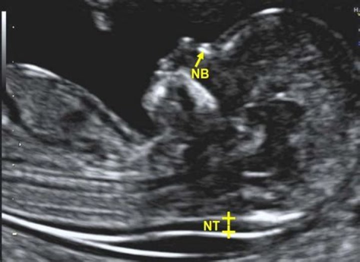
During the routine first trimester screening at 13 weeks of gestation, NT was measured at 3 mm. The normal range of NT for this age is 1.6-2.4 mm.
What is a NT scan at 13 weeks?
The NT ultrasound is done between 11 and 13 weeks, when baby’s nuchal translucency, the clear tissue located at the back of a developing baby’s neck, can be measured. An average NT measurement is around 2.18 millimeters.
What is an abnormal nuchal translucency measurement?
The fetal NT increases with gestational age/crown–rump length. Due to this the NT measurement may considered abnormal when it is above 3.0 mm, or above the 99th percentile for the gestational age.
What is the normal neck thickness 12 weeks?
At 12 weeks of gestational age, an “average” nuchal thickness of 2.18mm has been observed; however, up to 13% of chromosomally normal fetuses present with a nuchal translucency of greater than 2.5mm.Is 2.2 nuchal translucency normal?
A baby with an NT of 1.3mm is within the normal range. The baby with an NT of 2.9mm is also within the limit of normal range. As the NT increases, so does the chance of Down’s syndrome and other chromosomal conditions.
What if NT scan is not normal?
What if your NT results are abnormal? If your nuchal translucency screening or any other prenatal screening results indicate that your baby may be at an increased risk of having a genetic abnormality, your practitioner will likely suggest a diagnostic test like chorionic villus sampling (CVS) or amniocentesis.
How accurate is 12 week scan for Down's syndrome?
The first-trimester screening’s detection rate is approximately 96% for pregnancies in which the baby has Down syndrome and is somewhat higher for pregnancies with trisomy 13 or trisomy 18. 3 A nuchal translucency ultrasound can be performed without the bloodwork, but the detection rate is reduced to about 70%.
Can nuchal translucency be too low?
For a baby that is between 45 mm and 84 mm in size, an NT of less than 3.5mm is considered normal. An NT less than 1.3 mm is considered to be low-chance and an NT of 6 is considered high chance for Down’s syndrome and other potential chromosomal abnormalities.Is 0.9 mm nuchal translucency normal?
The average NT thickness was 1.7 mm (range from 0.9 mm to 13.4 mm). The NT was above the 95th centile of the normal range for the CRL in 75% (15 out of 20) of trisomy 21 pregnancies and in 64% (16 out of 25) pregnancies with other chromosomal abnormalities.
What if nuchal translucency is high?An increased NT has also been associated with a high risk of miscarriage or fetal death. This risk increases with increasing NT thickness, and miscarriage or fetal death may be preceded by cardiac failure symptoms such as fetal hydrops.
Article first time published onCan a thick nuchal translucency be normal?
Many healthy babies have thick nuchal folds. However, there is a higher chance for Down syndrome or other chromosome conditions when the nuchal fold is thick. There may also be a higher chance for rare genetic conditions.
What are soft markers for Down syndrome?
The most commonly studied soft markers of aneuploidy include a thickened nuchal fold, long bones shortening, mild fetal pyelectasis, echogenic bowel, echogenic intracardiac focus, FMF angle > 90 degrees, pathologic velocity of Ductus venosus and choroid plexus cyst.
How can you tell boy or girl from NT scan?
We can tell the sex of the baby at the 12 week scan by assessing the direction of the nub. This is something that can be identified on babies at this stage and if it points vertically then it is likely to be a boy. If it points horizontally then it is likely to be a girl.
What does a high NT measurement mean?
However, even if conventional karyotyping is normal, increased NT is a predictive value of adverse pregnancy outcome, because it is associated with several fetal malformations, congenital heart defects, genetic syndromes, intrauterine death and miscarriages; the majority of these structural anomalies are undetectable …
Can you have 12 week scan at 14 weeks?
The nuchal translucency scan is best done during the 12th week, but it can be done from 11 weeks and 3 days up until 14 weeks and your local NHS hospital will offer you an appointment to have this done at around this time.
What makes you high risk for Down's syndrome baby?
One factor that increases the risk for having a baby with Down syndrome is the mother’s age. Women who are 35 years or older when they become pregnant are more likely to have a pregnancy affected by Down syndrome than women who become pregnant at a younger age.
Can a 12 week scan show spina bifida?
From 12 weeks the spine can usually be seen clearly enough to rule out major cases of spina bifida. All of this information provides important reassurance. A more thorough evaluation of fetal anatomy will be done at 20-22 weeks gestation.
What is PAPP a normal range?
A Papp-A level more than or equal to 0.5 MOM is considered normal, while levels less than 0.5 MOM are marked as low.
Can a thick nuchal fold go away?
Natural course. An abnormally thickened nuchal fold or even a cystic hygroma may resolve, especially toward the third trimester; however, the risk of karyotypic abnormalities is not reduced.
How accurate are NT measurements?
How accurate is the nuchal translucency test? The NT scan alone will detect about 70 to 80 percent of babies with DS (depending on which study you look at). The detection rate for the NT scan plus a first-trimester blood test ranges from 79 to 90 percent. (Most tests will include both the NT scan and the blood test.)
What is a normal nuchal translucency measurement at 12 weeks NHS?
This is called nuchal translucency (NT). This collection of fluid normally measures less than 3.5mm between 11 and 14 weeks of pregnancy.
What is considered a low risk of Down syndrome?
The cut off is 1 in 150. This means that if your screening test results show a risk of between 1 in 2 to 1 in 150 that the baby has Down’s syndrome, this is classified as a higher risk result. If the results show a risk of 1 in 151 or more, this is classified as a lower risk result.
Can NT scan go wrong?
It’s important to remember that receiving an abnormal result from an NT scan doesn’t necessarily mean that your baby has a chromosome problem. Similarly, normal test results can’t guarantee that your baby won’t be born with Down syndrome. This test isn’t perfect. There’s a 5 percent false-positive rate.
Is nuchal translucency accurate?
The accuracy of a screening test is based on how often the test correctly finds a birth defect. The nuchal translucency test correctly finds Down syndrome in 64 to 70 out of 100 fetuses who have it. It misses Down syndrome in 30 to 36 out of 100 fetuses.
How common is a thick nuchal fold?
According to the practice bulletin concerning fetal aneuploidy screening published by the American Congress of Obstetricians and Gynecologists, the likely ratio (LR) for thickened nuchal fold (TNF) is 11 to 18.6.
What markers did your Down syndrome baby have?
Certain features detected during a second trimester ultrasound exam are potential markers for Down’s syndrome, and they include dilated brain ventricles, absent or small nose bone, increased thickness of the back of the neck, an abnormal artery to the upper extremities, bright spots in the heart, ‘bright’ bowels, mild …
Can you see Down syndrome on ultrasound?
An ultrasound can detect fluid at the back of a fetus’s neck, which sometimes indicates Down syndrome. The ultrasound test is called measurement of nuchal translucency. During the first trimester, this combined method results in more effective or comparable detection rates than methods used during the second trimester.
What does 2 soft markers mean?
If you were told that there were “soft markers” for Down syndrome during your level 2 ultrasound, you may be wondering what it means. A soft marker may indicate an increased likelihood of a chromosomal abnormality — but it’s simply not very reliable, especially considered outside of the bigger picture.
Can you tell gender at 13 week NT scan?
The accuracy of determining your baby’s gender increases with how far along you are in the pregnancy. The accuracy can vary from 70.3% at 11 weeks to 98.7% at 12 weeks, and 100% at 13 weeks. Eleven weeks is the earliest that sex determination can be carried out with an ultrasound using a method called the ‘nub theory’.
How accurate is a 14 week gender ultrasound?
Results: Results confirmed 100% accuracy in predictions made after 14 weeks gestation. The overall success rate in the first trimester group (11–14 weeks) was 75%. When excluding those scans where a prediction could not be made, success rates increased to 91%.
How often is a boy mistaken for a girl in an ultrasound?
The chances of an error with ultrasound are up to 5 percent, says Schaffir. An ultrasound can be between 95 to 99 percent accurate in determining sex, depending on when it’s done, how skilled the sonographer is and whether baby is in a position that shows the area between their legs. Mistakes can also be made.