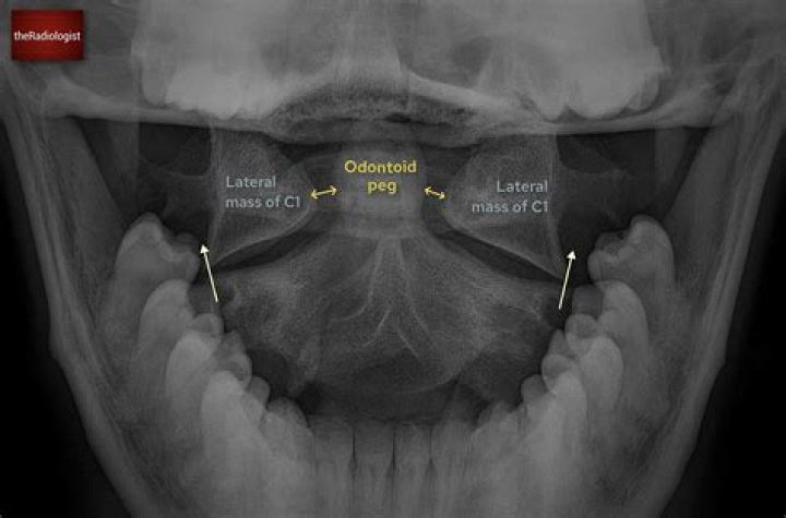
An open mouth view x-ray is a special view to visualize upper cervical spine problems especially C1 and C2 vertebrae. Routine anteroposterior cervical spine view shows the spine from C3 segment and is usually less helpful in diagnosing acute injuries.
Why do you open your mouth for neck x-ray?
The open mouth odontoid radiograph (x-ray) is used to assess for the presence of an upper cervical spine injury. Common injuries to the upper cervical spine include: Dens Fracture (i.e., C2 Odontoid Fracture) Jefferson’s Fracture (i.e., C1 Burst Fracture)
What is the central ray for AP open mouth projection?
C: For the open-mouth view, the patient is positioned in the same manner as for the supine AP projection; the head is straight, in the neutral position. With the patient’s mouth open as wide as possible, the CR is directed perpendicular to the midpoint of the open mouth.
What is an Odontoid view?
Odontoid view shows displacement of the lateral masses of C1, allowing distinction of this fracture from a simple fracture of the posterior neural arch of C1.When taking an AP cervical open mouth x-ray Where should the central ray be pointed?
This angle can and will vary between 5-20° depending on the position of the head. To project the intervertebral disc spaces open, the central ray should be directed perpendicular to the long axis of the vertebral column 3, 4.
Can a neck X-ray show a tumor?
If surgery of the cervical spine is required, an X-ray may be taken to plan for the surgery and to assess the post-operative results. A cervical spine X-ray also can give clues about an infection, tumor, or other abnormalities in the neck bones.
Would a neck X-ray show a tumor?
X-rays of the spine, neck, or back may be performed to diagnose the cause of back or neck pain, fractures or broken bones, arthritis, spondylolisthesis (the dislocation or slipping of 1 vertebrae over the 1 below it), degeneration of the disks, tumors, abnormalities in the curvature of the spine like kyphosis or …
On what vertebrae would you find the Odontoid process?
The odontoid process (also dens or odontoid peg) is a protuberance (process or projection) of the Axis (second cervical vertebra). It exhibits a slight constriction or neck, where it joins the main body of the vertebra.What does the odontoid do?
The odontoid process provides a pivot point — called an axis of motion — around which the skull and the first cervical vertebra (the atlas) rotate, twist and/or turn (these are really all the same thing.)
What causes odontoid fracture?Odontoid fractures occur as a result of trauma to the cervical spine. In younger patients, they are typically the result of high-energy trauma, which occurs as a result of motor vehicle or diving accidents.
Article first time published onWhat is Jefferson fracture?
A Jefferson fracture is a bone fracture of the vertebra C1. The vertebra C1 is a bony ring, with two wedge-shaped lateral masses, connected by relatively thin anterior and posterior arches and a transverse ligament. The lateral mass on vertebra C1, who is taller, is directed laterally.
What is the rule of Spence?
The Rule of Spence is a radiologic method to evaluate the likelihood of injury to the transverse atlantal ligament (TAL) on an open mouth AP (“peg”) radiograph.
Where is C1 and C2 on the spine?
The C1 and C2 vertebrae are the first two vertebrae at the top of the cervical spine. Together they form the atlantoaxial joint, which is a pivot joint.
Is your neck connected to your spine?
The neck is connected to the upper back through a series of seven vertebral segments. The cervical spine has 7 stacked bones called vertebrae, labeled C1 through C7.
Which sinus is not seen in waters view?
The sphenoid sinus was excluded, as it does not create a reliable image on open-mouth Waters radiographs. Fig. 1: The frontal and ethmoidal sinuses show mucosal thickening on the coronal view.
What is a thoracic X-ray?
A thoracic spine x-ray is an x-ray of the 12 chest (thoracic) bones (vertebrae) of the spine. The vertebrae are separated by flat pads of cartilage called disks that provide a cushion between the bones. This is the spine and the sacrum with the cervical (neck), thoracic (mid-back), and lumbar (lower back) vertebra.
How serious is an Odontoid fracture?
The overall complication rate was significantly higher in elderly (52.2% vs. 32.7%), with an in-hospital mortality of 34.8%. Elderly patients with a fracture of the odontoid are a high-risk group with a high morbidity /mortality.
What causes Anterolisthesis?
Anterolisthesis is often due to sudden blunt force or fractures. These can be the result of trauma typically experienced in an auto accident or a fall. Anterolisthesis can also develop over time through strenuous physical exercise, such as bodybuilding. Aging is another common cause of anterolisthesis.
What bones does the sacrum articulate with?
It articulates with the fifth lumbar vertebra, the os coxae, and the coccyx.
What are two features that cervical vertebrae have that other vertebrae do not?
The main anatomical characteristics of a typical cervical vertebra that separate it from other types of vertebrae are the small size, transverse foramina, saddle-shaped body, and bifid spinous process (Fig. 1.7.
What is spondylosis thesis?
Spondylolisthesis is a spinal condition that causes lower back pain. It occurs when one of your vertebrae, the bones of your spine, slips out of place onto the vertebra below it. Most of the time, nonsurgical treatment can relieve your symptoms. If you have severe spondylolisthesis, surgery is successful in most cases.
How do spinal nerves exit the vertebral column?
The spinal nerves leave the vertebral column through the intervertebral foraminae. Some spinal nerves are intermingled in plexuses, from which the peripheral nerves are formed, each nerve containing fibers from several spinal cord segments.
Can an Odontoid fracture heal on its own?
A stable fracture may “set” and heal itself. In an unstable fracture, the bone is more likely to move out of its normal position and alignment. Type II fractures are considered the least stable of the odontoid fractures. This makes them the most likely to require surgery.
How common are odontoid fractures?
Sometimes these fractures are missed or left untreated, and they can be associated with increased morbidity (disease) in older patients. Overall, odontoid fractures are the most common fracture of the C2 vertebra and can account for up to 15% of all cervical spine (neck) fractures.
How long does it take for a Jefferson fracture to heal?
What’s recovery like? If surgery is needed, recovery will likely take about 12 weeks. This is regardless of the type of surgery. If the fracture is minor, you may be able to get by with wearing a neck brace for six to eight weeks.
What is Brown Séquard syndrome?
Brown-Séquard syndrome is a rare spinal disorder that results from an injury to one side of the spinal cord in which the spinal cord is damaged but is not severed completely. It is usually caused by an injury to the spine in the region of the neck or back.
What is a Bennett fracture?
Bennett fracture is the most common fracture involving the base of the thumb. This fracture refers to an intraarticular fracture that separates the palmar ulnar aspect of the first metacarpal base from the remaining first metacarpal.
What is a Potts fracture?
A Pott’s fracture is a fracture affecting one or both of the malleoli. During activities such as landing from a jump (volleyball, basketball) or when rolling an ankle, a certain amount of stress is placed on the tibia and fibula and the ankle joint.
How long does it take for a c1 fracture to heal?
Isolated atlas fractures can be effectively managed with 8 to 12 weeks of external immobilization of the craniocervical junction [3]. Collar immobilization or cervical traction for this period of time is usually sufficient to allow for proper healing; however, the type of orthosis required varies [3, 20].
What is a C2 fracture?
The hangman’s fracture refers to a break in a bone known as C2, because it is the second bone down from the skull in your cervical (neck) vertebrae. A fracture can be a partial or complete break in a bone. The injury can also cause the C2 to move out of alignment with the bone right below it, known as the C3.
What is transverse ligament?
Description. The transverse ligament of the atlas (TLA) is a thick, strong band of approximately 20mm in length which arches across the ring of the atlas and maintains the odontoid process in contact with the anterior arch. It is concave in front, convex behind and broader at the middle than at the ends.